- Research
- Open access
- Published:
Maternal supplementation with n-3 fatty acids affects placental lipid metabolism, inflammation, oxidative stress, the endocannabinoid system, and the neonate cytokine concentrations in dairy cows
Journal of Animal Science and Biotechnology volume 15, Article number: 74 (2024)
Abstract
Background
The placenta plays a crucial role in supporting and influencing fetal development. We compared the effects of prepartum supplementation with omega-3 (n-3) fatty acid (FA) sources, flaxseed oil (FLX) and fish oil (FO), on the expression of genes and proteins related to lipid metabolism, inflammation, oxidative stress, and the endocannabinoid system (ECS) in the expelled placenta, as well as on FA profile and inflammatory response of neonates. Late-pregnant Holstein dairy cows were supplemented with saturated fat (CTL), FLX, or FO. Placental cotyledons (n = 5) were collected immediately after expulsion, and extracted RNA and proteins were analyzed by RT-PCR and proteomic analysis. Neonatal blood was assessed for FA composition and concentrations of inflammatory markers.
Results
FO increased the gene expression of fatty acid binding protein 4 (FABP4), interleukin 10 (IL-10), catalase (CAT), cannabinoid receptor 1 (CNR1), and cannabinoid receptor 2 (CNR2) compared with CTL placenta. Gene expression of ECS-enzyme FA-amide hydrolase (FAAH) was lower in FLX and FO than in CTL. Proteomic analysis identified 3,974 proteins; of these, 51–59 were differentially abundant between treatments (P ≤ 0.05, |fold change| ≥ 1.5). Top canonical pathways enriched in FLX vs. CTL and in FO vs. CTL were triglyceride metabolism and inflammatory processes. Both n-3 FA increased the placental abundance of FA binding proteins (FABPs) 3 and 7. The abundance of CNR1 cannabinoid-receptor-interacting-protein-1 (CNRIP1) was reduced in FO vs. FLX. In silico modeling affirmed that bovine FABPs bind to endocannabinoids. The FLX increased the abundance of inflammatory CD44-antigen and secreted-phosphoprotein-1, whereas prostaglandin-endoperoxide synthase 2 was decreased in FO vs. CTL placenta. Maternal FO enriched neonatal plasma with n-3 FAs, and both FLX and FO reduced interleukin-6 concentrations compared with CTL.
Conclusion
Maternal n-3 FA from FLX and FO differentially affected the bovine placenta; both enhanced lipid metabolism and modulated oxidative stress, however, FO increased some transcriptional ECS components, possibly related to the increased FABPs. Maternal FO induced a unique balance of pro- and anti-inflammatory components in the placenta. Taken together, different sources of n-3 FA during late pregnancy enhanced placental immune and metabolic processes, which may affect the neonatal immune system.
Background
In all pregnant mammals, the developing fetus requires substantial amounts of fatty acids (FAs) during late gestation to support rapid cellular growth and activity [1]. Generally, omega-3 FAs (n-3 FAs) have anti-inflammatory, pro-resolving, and anti-oxidative properties [2,3,4]. Furthermore, n-3 FAs play an essential role in cellular metabolism, energy storage, and maintenance of homeostasis. The n-3 FAs influence the abundance of lipid mediators [5, 6] and can alter the cell membrane composition [7]. The FA composition delivered to the fetus is largely determined by the maternal circulating levels, and the placenta was shown to preferentially transfer physiologically important long-chain polyunsaturated FA (LCPUFA), especially n-3 FAs [2]. The transfer of nutrients, including lipids, from mother to fetus, is influenced by multiple factors [8, 9]. The bovine placenta features chorionic villi that are arranged in a cotyledonary manner, generating a complex architecture of blood vessels. Based on the maternal-fetal blood communication and the cell composition, the bovine placenta is classified as epitheliochorial/synepitheliochorial. It is composed of trinucleate feto-maternal hybrid cells, which form multiple cell layers that completely separate the maternal and fetal vascular systems, thus limiting their communication [10,11,12,13]. Moreover, FA transfer between the maternal and the fetal interface depends on factors such as the size of the molecule, and the method of transfer, depending on whether it is via diffusion or some form of active or facilitated transport [10, 14]. Additionally, the expression of the FA binding proteins (FABPs) and FA transport proteins (FATPs), which have high affinities with arachidonic acid (AA; C20:4n-6) and docosahexaenoic acid (DHA; C22:6n-3), facilitate the transport of FAs from the maternal bloodstream to the fetal circulation [9, 15, 16].
During late-gestation and towards calving, the metabolic activity of placental cells is notably high, producing various growth factors and forming reactive oxygen species (ROS), as well as secreting inflammatory molecules, which play an important role both in promoting placental development and in detaching the placental maternal-fetal unit [17,18,19]. However, adverse conditions, such as nutritional stress, can lead to an increased release of inflammatory cytokines and the production of ROS, which consequently have a negative impact on placental functionality. This can also be induced by inadequate placental vascularization, as well as a reduction in the conversion ratios of LCPUFA and the supply of essential nutrients needed to ensure the proper development of organs and the health of the fetus [1, 20,21,22]. Specifically, within the placenta, n-3 FAs modulate energy metabolism and improve placental angiogenesis and fetal growth [23].
Supplementation with n-3 FAs can also modulate the activation of the endocannabinoid system (ECS) [24, 25] by reducing the omega-6 (n-6) FAs to the n-3 FA ratio in the diet [26]. The ECS, which consists of the endocannabinoid ligands, cannabinoid receptors, and enzymes that synthesize and degrade the endocannabinoids, is involved in regulating energy metabolism and immune function [27]. The endocannabinoids anandamide (AEA) and 2-arachidonoylglycerol (2-AG) are synthesized from AA, and are hydrolyzed back to AA, ethanolamine, and glycerol by the enzymes fatty acid amide hydrolase (FAAH) and monoglyceride lipase (MGLL), respectively [28, 29]. In our previous studies on peripartum dairy cows, we described the effects of supplementing various sources of n-3 FAs, such as flaxseed oil (FLX) and fish oil (FO), on the ECS and immune function in the cow. We observed that peripartum n-3 FA supplementation reduces the gene expression of the ECS components in white blood cells, liver, and the adipose tissue, lowered the expression of the molecule linked to signalization of the inflammatory pathway in white blood cells such as nuclear factor kappa B (NFκB), and decreased the blood percentage of immune cells that are active during the inflammatory process, including CD25+ T-regulatory, when compared with controls postpartum [24, 30, 31]. Thus, supplementation with n-3 FAs affected the immune response and the ECS components in peripartum dairy cows.
Recently, maternal n-3 FAs and methionine supplementation in ewes [32] were shown to decrease the gene expression of FA binding protein 4 (FABP4) in placental cotyledons, suggesting that maternal n-3 FAs affect placental lipid transfer. However, currently, there is no information on the effects of n-3 FAs on the placenta of dairy cows. Our objective was to investigate the effect of n-3 FA supplementation on lipid metabolism, inflammatory response, oxidative stress, and the ECS components in the placenta of dairy cows at calving as well as the effect of the cytokine concentration on the plasma of the neonates. To this end, we compared two sources of n-3 FAs: FLX and FO. FLX is a source of alpha-linolenic acid (ALA; C18:3n-3), which, within the body, can undergo limited conversion to form both DHA and eicosapentaenoic acid (EPA; C20:5n-3) [33, 34], whereas FO contains DHA and EPA [35]. We hypothesized that supplementation of peripartum dairy cows with n-3 FAs, rich in either EPA, DHA (FO), or in their precursor ALA (FLX), would modulate lipid metabolism by improving the transfer of n-3 FAs through the placenta to the fetus, reduce the ECS components in the placenta, and induce anti-inflammatory and anti-oxidative effects. We hypothesized that these changes may be beneficial for the placental function and may improve the inflammatory response in neonates.
Methods
Animals, treatments and experimental procedures
The experimental protocol for the study was approved by the Volcani Center Animal Care Committee (approval number IL 797/18), and it was performed in accordance with the relevant guidelines and regulations. The experiment was conducted at the Volcani Center experimental farm in Rishon Lezion, Israel. A detailed description of this experiment was provided in [30]. Briefly, 42 multiparous Holstein dairy cows, with a mean parity of 3.8 ± 1.4 (mean ± standard deviation), participated in the study during the winter season (November 2018–March 2019). The average temperature-humidity index (THI) was 61 ± 8, indicating conditions well within the thermoneutral zone for dairy cows [36]. The cows were group-housed from d 257 of pregnancy to d 60 postpartum in a shaded loose pen that was equipped with a real-time electronic individual feeding system. Each feeding station included an individual identification system (ID tag; SAE Afikim, Kibbutz Afikim, IL) that allowed each cow to enter a specific feeding station only and automatically recorded each meal. The cows were stratified according to milk yields during the first 60 d of the previous lactation, body weight at drying off, and parity. The prepartum dietary treatments began at d 257 of pregnancy as follows: (i) control group (CTL; n = 14), fed a basal dry cow diet supplemented with encapsulated saturated fatty acid (SFA) at 240 g/d/cow; (ii) FLX (n = 14), fed a basal diet supplemented at 300 g/d/cow, with encapsulated flaxseed oil providing ALA at 56.1 g/d; and (iii) FO (n = 14), fed a basal diet supplemented at 300 g/d/cow with encapsulated fish oil providing EPA at 5.8 g/d and DHA at 4.3 g/d. The total fat content of the CTL supplement was 99%, compared to 80% in the FLX and FO supplements; therefore, the amounts of the supplements differed between the groups in order to maintain similar fat contents in all diets. The fat supplements were specially prepared and supplied by SILA (Venice, Italy). The ingredients and the chemical compositions of the rations, as well as the profile of the main FAs in the supplements were previously presented in [30, 37]. The neonate calves were separated from their mothers at calving, and were provided individually with their mothers’ colostrum immediately after blood samples were collected post-calving. A diagram describing the experimental design, the treatments, biological samples collected, and laboratory analyses are presented (Additional file 1: Fig. S1).
Placenta collection
Placental tissues were collected immediately after the expulsion of the placenta postpartum. From each placenta, we dissected 4 or 5 samples (50 mg) of cotyledons that were stored immediately at −80 ºC for further RNA and protein extraction. In total, we collected 13 placenta samples during the experiment (3 CTL, 5 FLX, and 5 FO). Since we did not obtain sufficient placenta samples from the control cows, we collected 2 additional placenta samples from cows that were fed the basal dry cow diet (without SFA supplementation) during the winter season of the following year.
Placental RNA extraction and transcript expression
We analyzed the gene expression in 5 placenta samples from each dietary treatment. Total RNA extraction of placental tissue was performed using an Animal tissue RNA purification kit (#25700, NORGEN BioTek Corp, Ontario, Canada). The RNA purity was assessed using a NanoDrop One Microvolume UV-Vis Spectrophotometer (Thermo Scientific, Shoham, IL) with a 260/280 ratio of above 1.85. First-strand cDNA was generated using the RevertAid First Strand cDNA Synthesis Kit (#K1622, Thermo Fisher Scientific, Vilnius, Lithuania). Quantitative detection of specific mRNA transcripts was carried out by real-time PCR using a CFX Duet Real-Time PCR System (Bio-Rad Laboratories, Inc., Rishon LeZion, IL) with the SYBR Green PowerTrack™ Master Mix (#A46109, Applied BioSystems, MA, USA) and analyzed using Bio-Rad CFX Maestro Software. In placenta tissues we examined the transcription levels of genes related to lipid metabolism, inflammation, oxidative response, and ECS. The list of primers is presented in Table 1. The reference genes β-Actin (ACTB), glyceraldehyde-3-phosphate dehydrogenase (GAPDH), and ubiquitously expressed prefoldin like chaperone (UTX) were examined. NormFinder software suggested ACTB and UTX as the most stable housekeeping genes for the placenta samples. The relative quantity of each gene was normalized to the average transcription levels of the reference genes (2−∆∆Ct method) according to Livak et al. [38].
Placental protein extraction
The placental samples (15 mg) were ground in a BeadBug homogenizer (D1030-E, Benchmark Scientific, Sayreville, NJ, USA) with two 0.5-mm glass beads (#11079105, BioSpec, Bartlesville, OK, USA) and 1 mL lysis buffer [5% (w/v) sodium dodecyl sulfate (SDS, #L3771-100) in 100 mmol/L Tris-HCl buffer containing 1% (v/v) phenylmethylsulfonyl fluoride (PMSF, #P7626), 1% phosphatase inhibitor (#P5726), and 1% protease inhibitor (#P8340), all from Sigma-Aldrich, St. Louis, MO, USA]. After centrifugation at 20,000 × g for 15 min at 4 °C, the soluble fractions were collected and the protein concentrations were measured using a bicinchoninic acid (BCA) standard assay (9470BCAstand, Cyanogen, Bologna, Italy), then snap-frozen and stored at −80 °C.
Proteomic analysis
For proteomic analysis, we analyzed 3 placenta samples from each dietary treatment. From the controls, we selected the 3 samples that were available from the original experimental cohort, and in FLX and FO we randomly selected 3 samples from each treatment; overall, 9 placenta samples were subjected to mass spectrometry-based proteomic analysis. The samples were lysed with 5% SDS and digested with trypsin (#V528B, Promega, Madison, USA) using the S-trap method [39]. Samples were stored at −20 °C until further use.
LC/MS
Ultra Liquid Chromatography/Mass Spectrometry (ULC/MS)-grade solvents were used for all chromatographic steps. Each sample was loaded using nanoflow ultra performance liquid chromatography (UPLC) (nanoAcquity; Waters, Milford, MA, USA). The mobile phase consisted of (A) H2O + 0.1% formic acid (#06914144, Bio-Lab Ltd., Jerusalem, IL) and (B) acetonitrile (ACN, #0120502, Bio–Lab Ltd.) + 0.1% formic acid. The samples were desalted online using a Symmetry C18 trapping column (180 μm id, 20 mm length, 5 μm particle size; Waters). The peptides were then separated using a T3 HSS nano-column (75 μm id, 250 mm length, 1.8 μm particle size, Waters) at 0.35 µL/min. Peptides were eluted from the column into the mass spectrometer using the following gradient: 4%–29% B in 155 min, 29%–90% B in 5 min, maintained at 90% B for 5 min and then back to the initial conditions.
The Nano-UPLC was coupled online through a Nano-electrospray ionization mass spectrometry (NanoESI) emitter (10 μm tip; Fossil, Madrid, Span) to a Q Exactive HF mass spectrometer (Thermo Scientific, Massachusetts, USA). Data were acquired in data-dependent acquisition mode using the Top10 method. MS1 resolution was set to 120,000 (at 200 m/z), a mass range of 375–1,650 m/z, an automatic gain control (AGC) of 3e6, and the maximum injection time was set to 60 ms. MS2 was performed by isolation with the quadrupole, a width of 1.7 Th, 27 NCE, 15k resolution, an AGC target of 60 ms, and a dynamic exclusion of 45 s.
Proteomic data analysis
Raw data were processed with MetaMorpheus version 1.02 (available at https://github.com/smith-chem-wisc/MetaMorpheus). The following search settings were used: protease = trypsin, maximum missed cleavages = 2, minimum peptide length = 7, maximum peptide length = unspecified, initiator methionine behavior = Variable, fixed modifications = Carbamidomethyl on C, Carbamidomethyl on U, variable modifications = Oxidation on M, max mods per peptide = 2, max modification isoforms = 1,024, precursor mass tolerance = ± 5 parts per million (PPM), product mass tolerance = ± 20 PPM, and report peptide-spectrum match (PSM) ambiguity = True. The combined search database contained 37,704 non-decoy protein entries including 388 contaminant sequences. The proteins were quantified using the FlashLFQ method [40], embedded in MetaMorpheus. The quantitative comparisons were calculated using Perseus v1.6.2.3. Student’s t-test, after logarithmic transformation, was used to identify significant differences across the biological replica. Fold change (FC) was calculated based on the ratio of the geometric means of the case versus the control samples. Principal component analysis (PCA) revealed that one CTL sample was an outlier; therefore it was excluded from further analysis.
Bioinformatic analysis of proteomic data
Proteins with P ≤ 0.05 and a |FC| ≥ 1.5 were defined as significantly differentially abundant proteins (DAPs). Only DAPs with ≥ 1 unique peptides were analyzed using QIAGEN Ingenuity® Pathway Analysis (IPA) software (QIAGEN, Inc., https://digitalinsights.qiagen.com/IPA) to determine the most relevant pathways, functions, and networks altered by the dietary treatments. Volcano plots were plotted with Microsoft Excel and bubble plots were plotted using the SRplot free online platform [41].
In-silico docking studies of bovine FABPs with endocannabinoids
The FABPs link lipid metabolism and the ECS, since they serve as transporters of endocannabinoids within cells [42]; therefore, we aimed to determine whether bovine heart-type fatty acid binding protein (FABP3), epidermal-type fatty acid-binding protein (FABP5), and brain-type fatty acid binding protein (FABP7) bind to endocannabinoids by in silico modeling. To this end, FABP3 (Accession number: P10790), FABP5 (Accession number: P55052), and FABP7 (Accession number: Q09139) sequences were collected from the UNIPROT database using the Bos taurus model for modeling the protein structures. The endocannabinoids 2-AG, AEA and docosahexaenoyl ethanolamide (DHEA) were docked to target proteins using GOLD 3.0.1 software, a genetic algorithm that uses a strategy that covers three genetic operators such as migrations, mutations, and crossovers [43]. The compounds that docked into the active site of the target proteins were thoroughly studied by molecular mechanics calculations. The most energetically favorable conformation of each compound was identified and selected after docking. Each compound’s individual binding poses were studied, and interactions with the protein were calculated. A detailed description of the FABPs’ structure and docking modeling is shown in Additional file 3.
Blood samples collected from neonate calves, FA compositions in plasma and in placenta tissues and inflammatory ELISA analysis
Blood samples were collected from calves (n = 9 per treatment) immediately after calving, before colostrum offering. They were collected from the jugular vein into vacuum tubes containing lithium heparin (#BD367526, Becton Dickinson Systems, Cowley, UK). Plasma was separated following centrifugation at 1,500 × g for 20 min at 4 °C, and stored at −80 °C, pending analysis. The FA composition in the placenta and in the plasma of neonate calves was determined as described previously [44]. Briefly, the samples were saponified in a mixture of 60% potassium hydroxide (KOH; #UN1813, Merck, Darmstadt, DE) and ethanol (#UN1170, Bio-Lab Ltd., Jerusalem, IL), extracted with petroleum ether (#UN1268, Bio-Lab Ltd.), and methylated with 5% (v/v) sulfuric acid (#UN1830, Bio-Lab Ltd.) in methanol (#UN1230, Bio-Lab Ltd.). FA methyl esters were analyzed using a 7890N gas chromatograph (Agilent Technologies, Santa Clara, CA) equipped with a DB-23 capillary column (60 m × 0.25 mm × 0.25 μm; Agilent Technologies) and a flame ionization detector. The initial temperature of the column was set at 130 °C, which was increased by 6.5 °C/min to 170 °C, and then by 2.75 °C/min to 215 °C, and held at 215 °C for 18 min. Then, the temperature was increased to 230 °C at 40 °C/min for the remainder of the analysis. The carrier gas was hydrogen, flowing at a linear velocity of 1.6 mL/min; the injection volume was 1 µL.
Plasma interleukin 2 (IL-2) and interleukin 6 (IL-6) concentrations were determined using Bovine Duoset ELISA kits (#DY2465 and #DY8190, respectively; R&D Systems, Inc., Minneapolis, MN, USA). Plasma haptoglobin (HP) concentrations were examined using a haptoglobin bovine ELISA kit (#E-10HPT, ICL, Portland, OR, USA).
Statistical analysis
The placenta gene expression levels and the plasma variables from calves (IL-2, IL-6, and HP), as well as the FA composition from placenta or plasma were analyzed using a generalized linear model (GLM), using the following model: Yijk = µ + Ti + C(T)ij + Eijk, where Yijk = dependent variable, µ = overall mean, Ti = treatment effect (i = CTL, FLX, or FO), C(T)ij = cow or calf j nested in treatment i, and Eijk = random residual. The data were analyzed using 9.4 Statistical Analysis System software after verifying the normality via the Shapiro-Wilk PROC single variable residual. The data were shown as the mean ± standard error of the mean (SEM) and declared significant when it reached P ≤ 0.05, and a tendency at 0.05 < P < 0.10 by Tukey’s test. The percentages of long-chain n-3 FAs EPA (C20:5n-3), DHA (C22:6n-3) and their intermediate docosapentanoic acid (DPA, C22:5n-3) from the calves’ plasma were not normally distributed; therefore, we analyzed the frequency of the appearance of these FAs by PROC FREQ and they were declared significant when they reached P ≤ 0.05 by Fisher’s test.
Results
Effects of maternal n-3 FA supplementation on placental gene expression
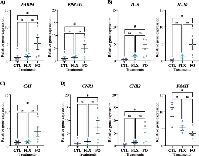
Several genes involved in lipid metabolism were examined; maternal FO supplementation increased the expression of the FABP4 gene, which is involved in FA transfer in the placenta, by 5-fold more than CTL (P = 0.05; Fig. 1A), with no difference observed between CTL and FLX. Additionally, there was a tendency towards higher gene expression of peroxisome proliferator-activated-receptor gamma (PPARG; P = 0.06; Fig. 1A) in the FO placenta, compared with CTL. On the other hand, the average relative expressions of sterol regulatory element binding transcription factor 1 (SREBP1; P = 0.51) and fatty acid synthase (FASN; P = 0.31) were similar between treatments (data not shown).
Expression levels of genes related to lipid metabolism, inflammation, oxidative stress, and ECS in placenta. (A) Lipid metabolism: FABP4 Fatty acid binding protein 4, PPARG Peroxisome proliferator activated receptor gamma; (B) Inflammation: IL-6 Interleukin 6, IL-10 Interleukin 10; (C) Oxidative stress: CAT Catalase; and (D) ECS components: CNR1 Cannabinoid receptor 1, CNR2 Cannabinoid receptor 2, FAAH Fatty acid amide hydrolase. Dairy cows at d 257 of pregnancy were divided into 3 nutritional groups supplemented with (i) CTL – encapsulated saturated fat, (ii) FLX – encapsulated flaxseed oil providing ALA, or (iii) FO – encapsulated fish oil providing EPA and DHA. Data represent the mean ± SEM. * P ≤ 0.05 and # P < 0.1 when comparing CTL, FLX, and FO treatments by Tukey’s test
In the analysis of the inflammation response genes, as shown in Fig. 1B, the FO placenta had higher gene expression of interleukin 10 (IL-10; P = 0.05) and a tendency for an increased expression of IL-6 (P = 0.06) compared with CTL, with no difference observed between FLX and CTL. No significant differences between treatments were observed regarding the placental gene expressions of interleukin 1b (IL-1b; P = 0.28), interleukin-6 receptor (IL6-R; P = 0.32), the toll-like receptor 4 (TLR4; P = 0.44), and tumor necrosis factor alpha (TNFα; P = 0.86; data not shown).
Among the oxidative stress-related genes, the expression of catalase (CAT; P = 0.04) was significantly higher in FO than in CTL (Fig. 1C). There were no differences in glutathione peroxidase 3 (GPX3; P = 0.17) and superoxide dismutase 1 (SOD1; P = 0.45) expressions between treatments (data not shown).
Furthermore, maternal FO supplementation led to a significant increase in the placental expression of the ECS-related genes cannabinoid receptor 1 (CNR1; P = 0.03) and cannabinoid receptor 2 (CNR2; P = 0.04) compared with CTL (Fig. 1D). Both n-3 FA treatments (FLX and FO) significantly decreased the placental gene expression of the ECS enzyme FAAH (P = 0.002) compared with CTL (Fig. 1D). No difference was observed between treatments regarding the gene expressions of the ECS enzymes MGLL (P = 0.20) and N-acyl phosphatidylethanolamine phospholipase D (NAPEPLD; P = 0.57; data not shown).
Effects of maternal n-3 FA supplementation on the placental proteome
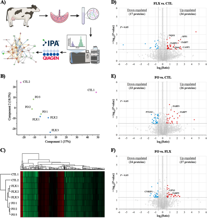
Overall, the proteomic analysis of the bovine placenta identified 3,974 proteins (Additional file 2). Of these, 51 proteins were differentially abundant (P ≤ 0.05 and |FC| ≥ 1.5) in FLX vs. CTL, whereas 59 were differential in FO vs. CTL, and 51 were differential in FO vs. FLX. Volcano plots illustrated that compared with CTL, the FLX treatment up-regulated 66.7% and down-regulated 33.3% of DAPs (Fig. 2D), whereas the FO treatment up-regulated 44.1% and down-regulated 55.9% of DAPs (Fig. 2E). Comparing DAPs in FO vs. FLX, 52.9% were up-regulated and 47.1% were down-regulated (Fig. 2F).
Proteomic analysis of the placenta of dairy cows supplemented pre-partum with n-3 FA. Placenta samples were collected immediately after delivery from dairy cows fed from d 257 of pregnancy with (i) CTL – encapsulated saturated fat, (ii) FLX – encapsulated flaxseed oil providing ALA, or (iii) FO – encapsulated fish oil providing EPA and DHA. (A) Work flow for the proteomic analysis; generated using BioRender.com. (B) Principal component analysis (PCA) of the placenta proteome; PCA analysis was used to assess the global integrity of the data and revealed that one CTL sample was an outlier, and therefore was excluded from further analysis; generated using Perseus v1.6.2.3. (C) Heat map analysis of the placental proteome: low peptide intensity is denoted in green, whereas high intensity is denoted in red. Each cow in the study was numbered and is represented in rows; generated using Perseus v2.0.11. (D–F) Volcano plot for the comparison between FLX vs. CTL (D), FO vs. CTL (E), and FO vs. FLX (F). P-value (≤ 0.05) is represented on the Y-axis and fold change (|FC| ≥ 1.5) is represented on the X-axis. Each dot represents one protein: red denotes up-regulated proteins, blue denotes down-regulated proteins
Of the DAPs, 2 proteins of the FABP family, FABP3 and FABP7, were significantly increased in FLX and FO compared with CTL. Interestingly, the proteomic data also revealed a higher abundance of the FABP5 (FC = 1.64, P = 0.04) in FO placenta vs. FLX. An additional protein implicated in the lipid metabolism was Caspase 2 (CASP2; FC = 5.04, P = 0.03), which was more abundant in FLX than in CTL. Furthermore, the n-3 FAs dietary treatments affected immune related proteins: FLX increased the abundance of CD44 antigen (CD44; FC = 2.04, P = 0.04) and secreted phosphoprotein 1 (SPP1; FC = 5.41, P = 0.02), whereas the prostaglandin-endoperoxide synthase 2 (PTGS2; FC = −1.97, P = 0.01) decreased in FO. The antioxidative protein NAD(P)H quinone dehydrogenase 1 (NQO1) increased in FLX, compared with CTL (FC = 1.51, P = 0.003), whereas GPX3 increased in FO, compared with FLX (FC = 1.67, P = 0.02). Another interesting protein that differed regarding FO vs. FLX was the ECS component CNR1-cannabinoid receptor-interacting protein 1 (CNRIP1), which was reduced in FO vs. FLX placenta (FC = −1.72, P = 0.02).
Top canonical pathways, functions and networks according to the differential placenta proteome
The DAPs (proteins with |FC| ≥ 1.5 and P ≤ 0.05) were analyzed using Qiagen’s Ingenuity® Pathway Analysis to identify the most relevant pathways, functions, and networks affected by FLX or FO dietary treatments. The main functions identified for DAP are presented in Additional file 1: Fig. S2. The most prominent biological function that changed between the treatments was regarding the levels of proteins with enzymatic functions: approximately 40% of DAPs in each comparison. Interestingly, FO supplementation resulted in a marked increase in the proportion of kinases and phosphatases among the differentially abundant enzymes, compared with FLX. Another important functional category identified in all the analyses was that of the proteins involved in transcription and translation regulation (Additional file 1: Fig. S2).
The top canonical pathways
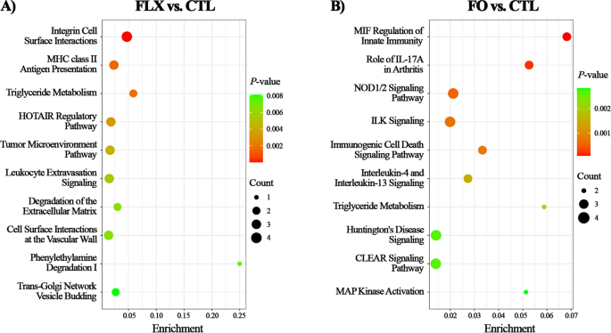
The top canonical pathways enriched in FLX vs. CTL placenta (Fig. 3A) were integrin cell surface interactions, major histocompatibility complex (MHC) class II antigen presentation, triglyceride metabolism, homeobox transcript antisense intergenic RNA (HOTAIR) regulatory pathway, and leukocyte extravasation signaling. Most pathways were mainly based on the differential abundance of FABP3, FABP7, CD44, and SPP1.
Top canonical pathway analysis according to differential proteome in placenta FLX or FO vs. CTL. Proteome analysis in placenta of dairy cows supplemented pre-partum with (i) CTL – encapsulated saturated fat, (ii) FLX – encapsulated flaxseed oil providing ALA, or (iii) FO – encapsulated fish oil providing EPA and DHA. (A) FLX vs. CTL and (B) FO vs. CTL. Enrichment (X-axis) is calculated by dividing the number of DAPs (|FC| ≥ 1.5) assigned to a particular pathway by the total number of molecules within that pathway. P-value (threshold of ≤ 0.05) is depicted by color scale. Plots were generated using SRplot [41]
In comparing FO to CTL (Fig. 3B), the top canonical pathways enriched were macrophage migration inhibitory factor (MIF) regulation of innate immunity, NOD1/2 signaling pathway, triglyceride metabolism, and mitogen-activated protein (MAP) kinase activation. Most pathways were based mainly on the differential abundance of FABP3, FABP7, PTGS2, and Nitric oxide synthase (NOS2). In comparing FO to FLX (Additional file 1: Fig. S3), the top canonical pathways annotated were RHO GTPases that activate protein kinases N (PKNs), triglyceride metabolism, triacylglycerol biosynthesis, and neutrophil degranulation. Most pathways were based mainly on the differential abundance of FABP5, phosphatidate phosphatase (LPIN2), and lysophosphatidylcholine acyltransferase 1 (LPCAT1).
Biological functions and networks
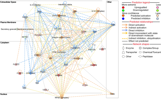
Among the top biological functions enriched in FLX vs. CTL (Table 2), those related to the FA metabolism (P = 0.0003), the concentration of lipid (P = 0.002), the synthesis of ROS (P = 0.02), the inflammation of organs (P = 0.000001), the inflammation of the absolute anatomical region (P = 0.0005), and the inflammation of the body cavity (P = 0.002) were most likely activated. In contrast, the biological functions related to the concentration of triacylglycerol (P = 0.004) were most likely inhibited. One of the main networks affected in FLX vs. CTL according to the DAP was lipid metabolism, molecular transport, and protein syntheses (Fig. 4). Some of the components related to the lipid metabolism network were CASP2, CD44, FABP3, FABP7, MAP2K1 protein (MAP2K1/2), NFKB2 protein (NFkB-complex), p38 mitogen-activated protein kinases (P38 MAPK), and platelet endothelial cell adhesion molecule (PECAM1).
Selected top scoring biological networks obtained for FLX vs. CTL using IPA analysis. Network ‘Lipid Metabolism, Molecular transport, and Protein synthesis’ includes the DAPs: CASP2 Caspase 2, CD44 Antigen CD44, FABP3 Heart-type fatty acid binding protein, FABP7 Brain-type fatty acid binding protein, brain, NFkB-complex NFKB2 protein, and P38 MAPK p38 mitogen-activated protein kinase. The image was generated using www.qiagen.com/ingenuity
Regarding FO vs. CTL (Table 2), the biological function of proteins related to FA metabolism (P = 0.01) was most likely activated, whereas functions related to the concentration of FAs (P = 0.02) were most likely inhibited. The top enriched molecular network regarding FO vs. CTL was cardiovascular system development and function, organ morphology, and organismal development (Additional file 1: Fig. S4A). Some of the components related to this network were caspase, CD59 molecule (CD59), FABP3, NOS2 and PTGS2.
Regarding FO vs. FLX (Table 2), the biological function affecting the concentration of lipid (P = 0.02) was most likely activated, whereas functions related to the synthesis of lipid (P = 0.03), production of ROS (P = 0.04), synthesis of ROS (P = 0.01), and inflammation of absolute anatomical region (P = 0.04) were most likely weakly inhibited. In comparing FO to FLX, one of the top networks affected was the cellular development, dermatological diseases and conditions, organismal injuries and abnormalities network (Additional file 1: Fig. S4B). The main components of this network are FABP5, NFkB complex, P38 MAPK, and complement factor H (CFH).
Docking studies of endocannabinoids with bovine FABPs
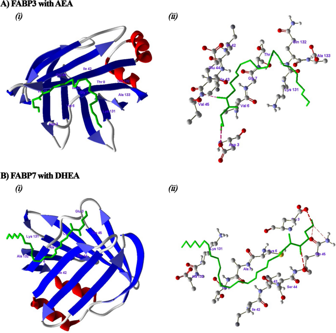
In-silico docking studies performed on bovine FABPs confirmed that FABP3 binds the n-6 series endocannabinoids AEA (4.16 kcal/mol, Fig. 5A) and 2-AG (6.35 kcal/mol, Additional file 3: Fig. S4) with good fitness scores. 2-AG and AEA also bind to FABP5 (5.90 kcal/mol and 3.55 kcal/mol, respectively) with good fitness scores (Additional file 3: Fig. S5). On the other hand, 2-AG and AEA had low fitness scores when binding to FABP7 (0.06 kcal/mol and 0.15 kcal/mol, respectively; Additional file 3: Fig. S6); however, as shown in Fig. 5B, FABP7 had a good binding score with the n-3 series endocannabinoid DHEA (10.43 kcal/mol).
In silico binding studies of the endocannabinoids with FABPs proteins from Bos taurus. (A) Docking studies of AEA with FABP3 and amino acids involved in hydrogen bonding with the AEA. (B) Docking studies of DHEA with FABP7 and amino acids involved in hydrogen bonding with the DHEA; hydrogen bonds were denoted by red dotted lines
Effects of maternal n-3 FA supplementation on FA composition in the placenta and plasma of neonate calves and on the inflammatory markers in neonate calves
First, we assessed the effects of maternal n-3 FA supplementation on the FA profile in the placenta tissues and the plasma of neonate calves. As shown in Table 3, in the placenta tissues, the average percentages of the n-3 FAs and the n-6/n-3 FA ratio remained similar across the treatments. On the other hand, in neonate calves, the plasma FA profile was affected by the maternal supplementation of the n-3 FAs; FO calves had a higher average percentage of total n-3 FAs (P = 0.02) compared with CTL (Table 4). Furthermore, a larger number of FO calves displayed increased percentages of plasma C20:5n-3 (EPA, P = 0.001), C22:6n-3 (DHA, P = 0.003), and their intermediate C22:5n-3 (DPA, P = 0.003), compared with both the CTL and FLX calves (Table 4).
Next, we evaluated the potential impact of maternal dietary n-3 FAs supplementation on the inflammatory status of neonatal calves by quantifying the plasma concentrations of several inflammatory markers on the day of calving before colostrum offering. A significant reduction of 14% and 22% in the average plasma concentrations of IL-6 was observed in the FLX and FO calves, respectively, compared with the CTL calves (P = 0.001; Table 5). On the other hand, there was no significant difference in IL-2 concentrations in both n-3 FAs treatments compared to CTL (Table 5); however, it was 22.8% higher in FLX than in the FO calves (P = 0.02). No differences were observed regarding the average concentrations of haptoglobin among the treatments (Table 5).
Discussion
Dietary n-3 FAs positively affect the physiological and reproductive properties in dairy cows [45]. In this work we aimed to investigate how maternal supplementation with different sources of n-3 FAs affects the genes and proteins related to the main physiological pathways in the placenta of dairy cows. We hypothesized that maternal n-3 FA supplementation would modulate lipid metabolism by improving the transfer of n-3 FAs through the placenta to the fetus, down-regulate ECS components, and have anti-inflammatory and anti-oxidative effects that may be beneficial for placental function and for the inflammatory response in neonates. Indeed, our proteomic approach, combined with gene expression data, showed that providing late-pregnant cows with FLX or FO, both sources of n-3 FAs, affects the bovine placenta via alterations in the expression patterns of proteins and genes related to these processes. Our findings of increased levels of inflammatory and ECS components in the FO placenta were unexpected, and will be discussed next.
Maternal n-3 FA supplementation affects the placental lipid metabolism
The fetus is dependent on the maternal supply of LCPUFAs; thus, the maternal FA composition is crucial for fetal growth and development, especially during late pregnancy, when DHA is particularly important for fetal brain development [46]. Placental cells are known to transfer the FAs selectively, with a preference for LCPUFAs, such as EPA and DHA. The selective uptake may involve intracellular metabolic channeling and a selective supply to the fetal circulation [2, 47, 48]. Several protein families were implicated in directional FA transport in the placenta: the membranal FATPs, FA translocase (FAT/CD36), and the cytoplasmic FABPs [2, 48, 49]. The uptake and accumulation of FAs are regulated by FABPs; FABP3, FABP4, FABP5, and FABP7 are expressed in cells with high FA requirements, such as the placenta [48, 50, 51]. In this study, we found that several FABPs were upregulated in FLX and/or the FO placenta; the abundances of FABP3 and FABP7 increased in both n-3 FAs groups, and these increases were related to the enriched pathway of triglyceride metabolism, whereas FABP4 gene expression was augmented by FO supplementation. Furthermore, FABP5 was higher in FO than in FLX placenta. FABPs have differential affinities to specific FAs: FABP5 binds to both DHA and AA, whereas FABP7 binds specifically to DHA [47, 52,53,54], and both FABP4 and FABP5 specifically increase the uptake of DHA [55,56,57,58]. Furthermore, although FABP3 was shown to bind preferentially to AA (n-6 FA) [54], it regulates both n-3 and n-6 FA transport in mouse trophoblasts [59]. Studies in bovine ovarian granulosa [60] and human placental cells [61] showed that the expression of FABP3, which may regulate the accumulation of triglycerides and the capacity of lipid transfer, can be influenced by the type of LCPUFA and the maternal health condition. In the present study, the plasma FA profile of neonate FO calves showed an increase in total n-3 FAs compared with CTL. We have previously demonstrated that the maternal plasma of FO cows was enriched with DHA and EPA [30]. Taken together, along with the increase in specific placental FABPs, we suggest that the increased maternal n-3 FAs may lead to increased FABPs in the placenta, which favors the trafficking of n-3 FAs to the fetus. The apparent lack of the effect of the dietary FA supplementation on the FA profile in placental cotyledons may stem from a highly efficient maternal-fetal transfer and/or rapid utilization of the FAs for placental functions.
Our proteomic data showed an increased abundance of CASP2 in the FLX placenta, compared with CTL. CASP2 is a protein in the apoptotic cascade, which was recently identified as a positive regulator of cholesterol and triacylglycerol homeostasis in human cells and mice [62, 63]. According to our bioinformatic analysis, CASP2 is involved in lipid concentration and triglyceride metabolism; it was demonstrated that the human CASP2 gene contains multiple sites that can be recognized by transcriptional regulators of the sterol regulatory element-binding protein (SREBP) family [62], which is involved in regulating pathways for cholesterol, triacylglycerol, and phospholipid synthesis [64]. In the placenta, the FAs are esterified into triglycerides and stored in droplets within the cells, after which they are transferred to the developing fetus [65]; thus, variations in the abundance of CASP2 may lead to modification of the cellular lipid levels, and specifically triacylglycerol [62, 63]. Therefore, our results suggest that maternal n-3 FA supplementation may modulate the metabolic pathway of triacylglycerol/lipid in the placenta by affecting the abundance of CASP2 protein.
Taken together, our results suggest that FO had a greater effect on the transfer of n-3 LCPUFAs to the fetus than did FLX. This transfer may be specifically mediated by modulating the FABPs in the placenta.
Maternal n-3 FA supplementation modulates ECS components in the placenta
Changing the n-3 to n-6 ratio in the diet is a well-known strategy to modulate the ECS [66]. We previously reported that in dairy cows, peripartum n-3 FA supplementation reduces the abundance of certain ECS components in the adipose tissue, liver, and white blood cells [24, 31]. However, in this study, in the placental tissue, FLX supplementation did not affect the gene expression of most of the examined ECS components, except for the FAAH gene. Intriguingly, the placenta from FO cows exhibited a higher gene expression of the primary membranal ECS receptors (CNR1 and CNR2), and a tendency toward increased gene expression of a secondary ECS nuclear receptor PPARG. Both the CNR1 and CNR2 receptors have important functions in reproductive organs, including the placenta [67, 68], since activation of the ECS receptors can affect both pregnancy progression and labor [29, 67] by controlling the action of endocannabinoids, cytokine release, and by modulating the mitochondrial activity [69, 70]. Within the cells, n-3 FA DHA and EPA can be converted to the endocannabinoids DHEA and eicosapentaenoyl ethanolamide (EPEA), which have a chemical structure similar to the endocannabinoid AEA; it was reported that DHEA and EPEA exhibit binding affinity and agonist activity on both CNR1 and CNR2 [71, 72]. Thus, the effects of FO on the ECS can be mediated by these n-3 FA-derived endocannabinoids. However, in the present study we did not measure the levels of endocannabinoids in placenta; therefore, further studies are required to investigate this possibility.
Interestingly, FABP3, FABP5, and FAB7 are also involved in the uptake, intracellular transport, and hydrolysis of endocannabinoids [51, 55, 73,74,75,76]. Since our data showed increased abundances of several FABPs in n-3 FA supplemented placentas, together with modulations in the ECS components, we investigated, for the first time in bovine, the in-silico binding of these Bos taurus FABPs with endocannabinoids. We found that FABP3 and FABP5 showed good fitness scores in the docking with both 2-AG and AEA, whereas bovine FABP7 had a good fitness score with DHEA. These findings may indicate an association between lipid metabolism and ECS within the bovine placenta. Furthermore, we observed a lower abundance of CNRIP1 protein in FO compared with FLX. CNRIP1 is involved in modulating part of the CNR1, thereby regulating cell signaling initiated by modulation of CNR1 [77]. However, the function of CNRIP1 in placental cells is not yet fully understood. To the best of our knowledge, this is the first study to identify CNRIP1 in the synepitheliochorial placenta, and there is a suggested link between n-3 FA supplementation and this ECS component.
Modulating the ECS can also affect inflammatory processes. In myoblast cell cultures, EPEA and DHEA decreased the gene expression of the inflammatory marker IL-6 [78]. However, in our study, the upregulation of the CNR in FO placenta coincided with a tendency to increase IL-6 expression. On the other hand, both FO and FLX placentas had reduced gene expression of FAAH compared with CTL. FAAH inhibition is considered to have anti-inflammatory effects [79, 80]. Taken together, we propose that maternal FO supplementation can stimulate placental ECS, which may be associated with activating the inflammatory and lipid metabolism pathways.
Maternal n-3 FA supplementation affects inflammation in the placenta and in calves
Within the placenta, pro-inflammatory cytokines are produced during parturition, most likely contributing to uterine contractions and the expulsion of the fetus [81, 82]. Note that we specifically sampled the cotyledons of the expelled placentas, namely, the maternal-fetal interface tissues post-detachment. When we examined the expression of inflammatory genes in the placental cotyledons, we found an increase in the expression of anti-inflammatory IL-10 and a tendency toward a higher expression of pro-inflammatory IL-6 in FO compared with CTL. In addition, an increase in the protein abundance of CD44 and SPP1, both associated with the activation of immune cells during inflammation [83], was found in FLX compared to CTL, and SPP1 tended to be increased in FO compared with CTL. Moreover, our analysis revealed an enrichment of the MHC class II antigen presentation and the leukocyte extravasation signaling pathways, as well as in the connection to inflammatory molecules, such as p38 MAPK and NFkB in the network, which are pro-inflammatory, in FLX vs. CTL [84]. In addition, in FO vs. CTL we found an enrichment in the MIF regulation of innate immunity, NOD1/2 signaling, and the MAP kinase activation pathways, which are associated with activation and control of the inflammatory process [85,86,87]. These findings indicate that maternal supplementation of n-3 FAs mostly upregulates the inflammatory process in the expelled placenta, which contradicts the well-described anti-inflammatory effects of n-3 FAs in other tissues. However, our analyses were conducted specifically on placental cotyledons, revealing a specific localized pro-inflammatory response. This observation aligns with the findings of Peng et al. [23], who demonstrated that EPA supplementation can induce a distinctive local pro-inflammatory effect in placenta from mice. However, in contrast to the putative upregulation of the inflammatory processes in the placenta, the proteomic data in FO vs. CTL showed a decreased abundance of PTGS2, encoding the enzyme cyclooxygenase-2, which regulates the synthesis of pro-inflammatory prostaglandins [88, 89]. The lower abundance of PTGS2 may indicate the anti-inflammatory effects of FO, which is in line with the increased gene expression of IL-10, thus overall, suggesting that FO has both pro- and anti-inflammatory effects on the bovine placenta.
In the present study we also investigated the effects of maternal n-3 FA supplementation on inflammatory markers in the neonate calves. We found that both FLX and FO reduced the plasma concentrations of IL-6 in neonates, which is in accordance with the well-described anti-inflammatory effects of n-3 FAs. Although the placental synepteliochorial does not allow the transfer of large molecules, such as immunoglobulins and lipid-insoluble molecules from the maternal blood flow to the fetus [12, 90], based on our results, it is possible that during late pregnancy, the maternal diet can influence the inflammatory response of neonates before the ingestion of colostrum, by facilitating placental transfer of n-3 FAs that modulate the release of cytokines, thus, highlighting the role of the placenta in the immune status of neonates during their first hours of life.
Maternal n-3 FA supplementation affects the placental oxidative stress response
The placental cells exhibit a high level of mitochondrial activity, which leads to the production of ROS [17] and increased oxidative stress [91]. Our proteomic data indicated that ROS synthesis was altered significantly in FLX placenta compared with CTL. In addition, we identified the up-regulation of CAT expression in FO placentas, whereas the expressions of GPX3 and SOD1 were similar across the treatments. In rats, n-3 FA supplementation increases the oxidative stress response in placental tissue, along with a positive correlation with the gestational quality [92]. Dietary n-3 PUFAs, and specifically DHA, were shown to act as activators of the Nrf2 antioxidant pathway [93,94,95,96]. Nrf2 is a transcriptional activator that regulates the expression of antioxidant genes including SOD1, NQO1, CAT, and GPX3 [97]. Indeed, we found that the abundance of NQO1 increased in the placental cotyledon cells of the FLX group. The NQO1 is a cytoplasmic protein that plays a key role in protecting against oxidative stress by stabilizing various proteins and preventing the reduction of a single electron that leads to the production of ROS [8]. We found that in FLX vs. CTL, NQO1 was most likely activated in biological functions related to ROS synthesis. Interestingly, although the analysis of the proteomic data did not reveal a significant impact on the ROS-related functions in the FO placenta, we did find an increased gene expression of the antioxidant enzyme CAT in FO compared with CTL. In a study with diabetic rats, supplementation with FO increased the gene expression of CAT, which is in agreement with our findings [98]. In the present study, there was a decrease in the gene expression of the ECS enzyme FAAH in both FO and FLX, compared with CTL. FAAH degrades AEA into ethanolamine and AA. AA, besides constituting a high percentage of the membrane phospholipids in cells, also serves as a precursor for the synthesis of prostaglandins-ethanolamids or ‘prostamides’, such as prostamide-E2 (PME2) [5, 89]. PME2 exhibits proapoptotic action and can lead to the production of ROS [68, 89]; therefore, reduction of FAAH may lessen oxidative stress.
In comparing the FO and FLX treatments, two functions associated with the production and synthesis of ROS were most likely inhibited: One of the proteins assigned to this function was GPX3, a member of the enzyme antioxidant family whose main biological role is to minimize oxidative damage by catalyzing the removal of lipid peroxides, which are products of ROS activity [99]. Indeed, GPX3 abundance was upregulated in FO compared with FLX. In sheep, Garrel et al. [100] suggested that GPX may be the major enzyme in defending against ROS in the fetus-placental unit. Overall, the increase in the oxidative stress response in the n-3 FA supplemented placental tissues is important to preserve gestational function [92]. An important consideration in interpreting our results is the specific highly- specialized tissue examined and the timing of the sample collection, which displays a physiological increase in the oxidative processes. Based on our results indicating that several oxidative stress components at the transcriptional and post-transcriptional levels were stimulated both in FLX and in FO, we suggest that maternal supplementation of n-3 FAs can have antioxidant effects on the bovine placenta.
Conclusions
Maternal n-3 FA from FLX and FO differentially affected the bovine placenta; both enhanced lipid metabolism and modulated oxidative stress; however, FO increased some transcriptional ECS components, possibly related to the increased FABPs. Maternal FO induced a unique balance of pro- and anti-inflammatory components in the placenta. Both n-3 FA supplementations altered the inflammatory markers in neonatal blood. Taken together, different sources of n-3 FA during late pregnancy affected placental immune and metabolic processes, which may affect the neonatal immune system.
Availability of data and materials
The proteomic dataset generated and/or analyzed during the current study is available in the Supplementary files.
Abbreviations
- 2-AG:
-
2-arachidonoylglycerol
- AA:
-
Arachidonic acid
- ACTB:
-
β-Actin
- AEA:
-
Anandamide
- AGC:
-
Automatic gain control
- ALA:
-
Alpha-linolenic acid
- CASP2:
-
Caspase 2
- CAT:
-
Catalase
- CD44:
-
CD44 antigen
- CD59:
-
CD59 molecule
- CFH:
-
Complement factor H
- CNR1:
-
Cannabinoid receptor 1
- CNR2:
-
Cannabinoid receptor 2
- CNRIP1:
-
CNR1-cannabinoid receptor interacting protein 1
- CTL:
-
Control group
- DAP:
-
Differently abundant protein
- DHA:
-
Docosahexaenoic acid
- DHEA:
-
Docosahexaenoyl ethanolamide
- DPA:
-
Docosapentanoic acid
- ECS:
-
Endocannabinoid system
- ELISA:
-
Enzyme-linked immunosorbent assays
- EPA:
-
Eicosapentaenoic acid
- EPEA:
-
Eicosapentaenoyl ethanolamide
- FA :
-
Fatty acid
- FAAH:
-
Fatty acid amide hydrolase
- FABP3:
-
Heart-type fatty acid binding protein
- FABP4:
-
Fatty acid binding protein 4
- FABP5:
-
Epidermal-type fatty acid-binding protein
- FABP7:
-
Brain-type fatty acid binding protein
- FABP:
-
Fatty acid binding protein
- FASN:
-
Fatty acid synthase
- FAT/CD36:
-
Fatty acid translocase
- FATPs:
-
Fatty acid transport proteins
- FC:
-
Fold change
- FLX:
-
Flaxseed oil
- FO:
-
Fish oil
- GAPDH:
-
Glyceraldehyde-3-phosphate dehydrogenase
- GLM:
-
Generalized linear model
- GPX3:
-
Glutathione peroxidase 3
- HP:
-
Haptoglobin
- HOTAIR:
-
Homeobox transcript antisense intergenic RNA
- IL-1b:
-
Interleukin 1b
- IL-2:
-
Interleukin 2
- IL-6:
-
Interleukin 6
- IL6-R:
-
Interleukin-6 receptor
- IL-10:
-
Interleukin 10
- IPA:
-
Ingenuity® pathway analysis
- KOH:
-
Potassium hydroxide
- LC/MS:
-
Liquid chromatography tandem mass spectrometry
- LCPUFA:
-
Long-chain polyunsaturated fatty acids
- LPCAT1:
-
Lysophosphatidylcholine acyltransferase 1
- LPIN2:
-
Phosphatidate phosphatase
- MAP:
-
Mitogen-activated protein
- MAP2K1/2:
-
MAP2K1 protein
- MGLL:
-
Monoglyceride lipase
- MIF:
-
Macrophage migration inhibitory factor
- MHC:
-
Major histocompatibility complex
- n-3 FA :
-
Omega-3 fatty acid
- n-6 FA :
-
Omega-6 fatty acid
- NAPEPLD:
-
N-acyl phosphatidylethanolamine phospholipase D
- NCE:
-
Normalized collision energy
- NFkB :
-
Nuclear factor kappa B
- NFkB-complex:
-
NFKB2 protein
- NanoESI:
-
Nano-electrospray ionization mass spectrometry
- NOS2:
-
Nitric oxide synthase
- NQO1:
-
NAD(P)H quinone dehydrogenase 1
- P38 MAPK:
-
p38 mitogen-activated protein kinases
- PCA :
-
Principal component analysis
- PCR :
-
Polymerase chain reaction
- PECAM1:
-
Platelet endothelial cell adhesion molecule
- PKNs:
-
Protein kinases N
- PME2:
-
Prostamide-E2
- PMSF:
-
Phenylmethylsulfonyl fluoride
- PPARG:
-
Proliferator-activated- receptor gamma
- PPM:
-
Parts per million
- PSM:
-
Peptide-spectrum match
- PTGS2:
-
Prostaglandin–endoperoxide synthase 2
- RNA :
-
Ribonucleic acid
- ROS:
-
Reactive oxygen species
- RT-PCR:
-
Reverse transcription polymerase chain reaction
- SDS:
-
Sodium dodecyl sulfate
- SEM:
-
Standard error of the mean
- SFA:
-
Saturated fatty acid
- SOD1:
-
Superoxide dismutase1
- SPP1:
-
Secreted phosphoprotein 1
- SREBPs:
-
Sterol regulatory element-binding protein
- SREBP1:
-
Sterol regulatory element binding transcription factor 1
- Th:
-
Thompsons
- THI:
-
Temperature-humidity index
- TLR4:
-
Toll like receptor 4
- TNFα:
-
Tumor necrosis factor alpha
- UPLC:
-
Ultra Performance Liquid Chromatography
- ULC/MC:
-
Ultra Liquid Chromatography/Mass Spectrometry
- UTX:
-
Ubiquitously expressed prefoldin like chaperone
- ∆∆Ct:
-
Delta-delta-cycle threshold
References
Duttaroy AK, Basak S. Maternal dietary fatty acids and their roles in human placental development. Prostaglandins Leukot Essent Fat Acids. 2020;155:102080.
Jones ML, Mark PJ, Waddell BJ. Maternal dietary omega-3 fatty acids and placental function. Reproduction. 2014;54:2247–54.
Goff JP, Horst RL. Physiological changes at parturition and their relationship to metabolic disorders. J Dairy Sci. 1997;80:1260–8.
Lessard M, Gagnon N, Godson DL, Petit HV. Influence of parturition and diets enriched in n-3 or n-6 polyunsaturated fatty acids on immune response of dairy cows during the transition period. J Dairy Sci. 2004;87:2197–210.
Calder PC. Omega-3 fatty acids and inflammatory processes. Nutrients. 2010;2:355–74.
Calder PC. Fatty acids: long-chain fatty acids and inflammation. Proc Nutr Soc. 2012;71:284–9.
Sokoła-Wysoczańska E, Wysoczański T, Wagner J, Czyż K, Bodkowski R, Lochyński S, et al. Polyunsaturated fatty acids and their potential therapeutic role in cardiovascular system disorders-a review. Nutrients. 2018;10:1561.
Preethi S, Arthiga K, Patil AB, Spandana A, Jain V. Review on NAD(P)H dehydrogenase quinone 1 (NQO1) pathway. Mol Biol Rep. 2022;49:8907–24.
Larqué E, Demmelmair H, Gil-Sánchez A, Prieto-Sánchez MT, Blanco JE, Pagán A, et al. Placental transfer of fatty acids and fetal implications. Am J Clin Nutr. 2011;94:1908–13.
Furukawa S, Kuroda Y, Sugiyama A. A comparison of the histological structure of the placenta in experimental animals. J Toxicol Pathol. 2014;27:11–8.
Haeger JD, Hambruch N, Pfarrer C. The bovine placenta in vivo and in vitro. Theriogenology. 2016;86:306–12.
Wooding FBP. The ruminant placental trophoblast binucleate cell: an evolutionary breakthrough. Biol Reprod. 2022;107:705–16.
Wooding FBP. The synepitheliochorial placenta of ruminants: Binucleate cell fusions and hormone production. Placenta. 1992;13:101–13.
Battaglia FC, Meschia G. Fetal nutrition. Annu Rev Nutr. 1988;8:43–61.
Campbell FM, Gordon MJ, Dutta-Roy AK. Placental membrane fatty acid-binding protein preferentially binds arachidonic and docosahexaenoic acids. Life Sci. 1998;63:235–40.
Roque-Jimenez JA, Oviedo-Ojeda MF, Whalin M, Lee-Rangel HA, Relling AE. Eicosapentaenoic and docosahexaenoic acid supplementation during early gestation modified relative abundance on placenta and fetal liver tissue mRNA and concentration pattern of fatty acids in fetal liver and fetal central nervous system of sheep. PLoS One. 2020;15:e0235217.
Walker OS, Ragos R, Wong MK, Adam M, Cheung A, Raha S. Reactive oxygen species from mitochondria impacts trophoblast fusion and the production of endocrine hormones by syncytiotrophoblasts. PLoS One. 2020;15:e0229332.
Pereira RD, De Long NE, Wang RC, Yazdi FT, Holloway AC, Raha S. Angiogenesis in the placenta: the role of reactive oxygen species signaling. Biomed Res Int. 2015;2015:814543.
Hirayama H, Sakumoto R, Koyama K, Yasuhara T, Hasegawa T, Inaba R, et al. Placentomes at spontaneous and induced parturition. J Reprod Dev. 2020;66:49–55.
Cetin I, Alvino G. Intrauterine growth restriction: implications for placental metabolism and transport. Rev Placenta. 2009;30:77–82.
Karowicz-Bilinska A, Kȩdziora-Kornatowska K, Bartosz G. Indices of oxidative stress in pregnancy with fetal growth restriction. Free Radic Res. 2007;41:870–3.
White MR, Yates DT. Dousing the flame: reviewing the mechanisms of inflammatory programming during stress-induced intrauterine growth restriction and the potential for ω-3 polyunsaturated fatty acid intervention. Front Physiol. 2023;14:1250134.
Peng JJ, Zhou Y, Hong Z, Wu Y, Cai A, Xia M, et al. Maternal eicosapentaenoic acid feeding promotes placental angiogenesis through a Sirtuin-1 independent inflammatory pathway. BBA-Molecular Cell Biol Lipids. 2019;1864:147–57.
Kra G, Daddam JR, Moallem U, Kamer H, Mualem B, Levin Y, et al. Alpha-linolenic acid modulates systemic and adipose tissue-specific insulin sensitivity, inflammation, and the endocannabinoid system in dairy cows. Sci Rep. 2023;13:5280.
Kim J, Li Y, Watkins BA. Fat to treat fat: emerging relationship between dietary PUFA, endocannabinoids, and obesity. Prostaglandins Other Lipid Mediat. 2013;104–105:32–41.
Zachut M, Tam J, Contreras GA. Modulating immunometabolism in transition dairy cows: the role of inflammatory lipid mediators. Anim Front. 2022;12:37–45.
Myers MN, Zachut M, Tam J, Contreras GA. A proposed modulatory role of the endocannabinoid system on adipose tissue metabolism and appetite in periparturient dairy cows. J Anim Sci Biotechnol. 2021;12:21.
Watson JE, Kim JS, Das A. Emerging class of omega-3 fatty acid endocannabinoids & their derivatives. Prostaglandins Other Lipid Mediat. 2019;143:106337.
Dirandeh E, Ansari-Pirsaraei Z, Deldar H, Shohreh B, Ghaffar J. Endocannabinoid system and early embryonic loss in holstein dairy cows. Anim Sci Pap Rep. 2020;38:135–44.
Kra G, Nemes-Navon N, Daddam JR, Livshits L, Jacoby S, Levin Y, et al. Proteomic analysis of peripheral blood mononuclear cells and inflammatory status in postpartum dairy cows supplemented with different sources of omega-3 fatty acids. J Proteom. 2021;246:104313.
Kra G, Daddam JR, Moallem U, Kamer H, Kočvarová R, Nemirovski A, et al. Effects of omega-3 supplementation on components of the endocannabinoid system and metabolic and inflammatory responses in adipose and liver of peripartum dairy cows. J Anim Sci Biotechnol. 2022;13:114.
Rosa Velazquez M, Batistel F, Pinos Rodriguez JM, Relling AE. Effects of maternal dietary omega-3 polyunsaturated fatty acids and methionine during late gestation on fetal growth, DNA methylation, and mRNA relative expression of genes associated with the inflammatory response, lipid metabolism and DNA methylation in placenta and offspring’s liver in sheep. J Anim Sci Biotechnol. 2020;11:111.
Lane K, Derbyshire E, Li W, Brennan C. Bioavailability and potential uses of vegetarian sources of omega-3 fatty acids: a review of the literature. Crit Rev Food Sci Nutr. 2014;54:572–9.
Takic M, Pokimica B, Petrovic-Oggiano G, Popovic T. Effects of dietary α-linolenic acid treatment and the efficiency of its conversion to eicosapentaenoic and docosahexaenoic acids in obesity and related diseases. Molecules. 2022;27:4471.
Calder PC, Yaqoob P, Thies F, Wallace FA, Miles EA. Fatty acids and lymphocyte functions. Br J Nutr. 2002;87:S31–48.
Silanikove N, Koluman DN. Impact of climate change on the dairy industry in temperate zones: predications on the overall negative impact and on the positive role of dairy goats in adaptation to earth warming. Small Rumin Res. 2015;123:27–34.
Moallem U, Shafran A, Zachut M, Dekel I, Portnick Y, Arieli A. Dietary α-linolenic acid from flaxseed oil improved folliculogenesis and IVF performance in dairy cows, similar to eicosapentaenoic and docosahexaenoic acids from fish oil. Reproduction. 2013;146:603–14.
Livak KJ, Schmittgen TD. Analysis of relative gene expression data using real-time quantitative PCR and the 2–∆∆CT method. Methods. 2001;25:402–8.
Elinger D, Gabashvili A, Levin Y. Suspension trapping (S-Trap) is compatible with typical protein extraction buffers and detergents for bottom-up proteomics. J Proteome Res. 2019;18:1441–5.
Millikin RJ, Solntsev SK, Shortreed MR, Smith LM. Ultrafast peptide label-free quantification with FlashLFQ. J Proteome Res. 2018;17:386–91.
Tang D, Chen M, Huang X, Zhang G, Zeng L, Zhang G, et al. SRplot: a free online platform for data visualization and graphing. PLoS One. 2023;18:e0294236.
Hamilton J, Roeder N, Richardson B, Hammond N, Sajjad M, Yao R, et al. Unpredictable chronic mild stress differentially impacts resting brain glucose metabolism in fatty acid-binding protein 7 deficient mice. Psychiatry Res-Neuroimaging. 2022;323:111486.
Kra G, Daddam JR, Moallem U, Kamer H, Ahmad M, Nemirovski A, et al. Effects of environmental heat load on endocannabinoid system components in adipose tissue of high yielding dairy cows. Animals. 2022;12:795.
Moallem U, Folman Y, Bor A, Arav A, Sklan D. Effect of calcium soaps of fatty acids and administration of somatotropin on milk production, preovulatory follicular development, and plasma and follicular fluid lipid composition in high yielding dairy cows. J Dairy Sci. 1999;82:2358–68.
Moallem U. Invited review: roles of dietary n-3 fatty acids in performance, milk fat composition, and reproductive and immune systems in dairy cattle. J Dairy Sci. 2018;101:8641–61.
Bernardi JR, Escobar RDS, Ferreira CF, Silveira PP. Fetal and neonatal levels of omega-3: effects on neurodevelopment, nutrition, and growth. Sci World J. 2012;2012:1–8.
Johnsen GM, Weedon-Fekjær MS, Tobin KAR, Staff AC, Duttaroy AK. Long-chain polyunsaturated fatty acids stimulate cellular fatty acid uptake in human placental choriocarcinoma (BeWo) cells. Placenta. 2009;30:1037–44.
Duttaroy AK. Transport of fatty acids across the human placenta: a review. Prog Lipid Res. 2009;48:52–61.
Wu Z, Hu G, Zhang Y, Ao Z. IGF2 may enhance placental fatty acid metabolism by regulating expression of fatty acid carriers in the growth of fetus and placenta during late pregnancy in pigs. Genes (Basel). 2023;14:872.
Larqué E, Krauss-Etschmann S, Campoy C, Hartl D, Linde J, Klingler M, et al. Docosahexaenoic acid supply in pregnancy affects placental expression of fatty acid transport proteins. Am J Clin Nutr. 2006;84:853–61.
Wang C, Mu T, Feng X, Zhang J, Gu Y. Study on fatty acid binding protein in lipid metabolism of livestock and poultry. Res Vet Sci. 2023;158:185–95.
Maximin E, Langelier B, Aïoun J, Al-Gubory KH, Bordat C, Lavialle M, et al. Fatty acid binding protein 7 and n-3 poly unsaturated fatty acid supply in early rat brain development. Dev Neurobiol. 2016;76:287–97.
Murphy EJ, Owada Y, Kitanaka N, Kondo H, Glatz JFC. Brain arachidonic acid incorporation is decreased in heart fatty acid binding protein gene-ablated mice. Biochemistry. 2005;44:6350–60.
Tokuyama S, Nakamoto K. Pain as modified by polyunsaturated fatty acids. In: Watson RR, Meester FD, editors. Omega-3 fatty acids in brain and neurological health. Waltham: Elsevier Academic Press; 2014. p. 131–46.
Liu JW, Almaguel FG, Bu L, De Leon DD, De Leon M. Expression of E-FABP in PC12 cells increases neurite ex- tension during differentiation: involvement of n-3 and n-6 fatty acids. Int Soc Neurochem. 2008;105:2015-29.
Lei C, Fan B, Tian J, Li M, Li Y. PPARγ regulates FABP4 expression to increase DHA content in golden pompano (Trachinotus ovatus) hepatocytes. Br J Nutr. 2022;127:3–11.
Pan Y, Short JL, Choy KHC, Zeng AX, Marriott PJ, Owad Y, et al. Fatty acid-binding protein 5 at the blood–brain barrier regulates endogenous brain docosahexaenoic acid levels and cognitive function. J Neurosci. 2016;36:11755–67.
Pan Y, Scanlon MJ, Owada Y, Yamamoto Y, Porter CJH, Nicolazzo JA. Fatty acid-binding protein 5 facilitates the blood-brain barrier transport of docosahexaenoic acid. Mol Pharm. 2015;12:4375–85.
Islam A, Kagawa Y, Sharifi K, Ebrahimi M, Miyazaki H, Yasumoto Y, et al. Fatty acid binding protein 3 is involved in n-3 and n-6 PUFA transport in mouse trophoblasts. J Nutr. 2014;144:1509–16.
Zhang N, Wang L, Luo G, Tang X, Ma L, Zheng Y, et al. Arachidonic acid regulation of intracellular signaling pathways and target gene expression in bovine ovarian granulosa cells. Animals. 2019;9:374.
Mishra JS, Zhao H, Hattis S, Kumar S. Elevated glucose and insulin levels decrease DHA transfer across human trophoblasts via SIRT1-dependent mechanism. Nutrients. 2020;12:1271.
Logette E, Le Jossic-Corcos C, Masson D, Solier S, Sequeira-Legrand A, Dugail I, et al. Caspase-2, a novel lipid sensor under the control of sterol regulatory element binding protein 2. Mol Cell Biol. 2005;25:9621–31.
Wilson CH, Nikolic A, Kentish SJ, Keller M, Hatzinikolas G, Dorstyn L, et al. Caspase-2 deficiency enhances whole-body carbohydrate utilisation and prevents high-fat diet-induced obesity. Cell Death Dis. 2017;8:e3136.
Eberlé D, Hegarty B, Bossard P, Ferré P, Foufelle F. SREBP transcription factors: master regulators of lipid homeostasis. Biochimie. 2004;86:839–48.
Lewis RM, Desoye G. Placental lipid and fatty acid transfer in maternal overnutrition. Ann Nutr Metab. 2017;70:228–31.
Zachut M. Letter to the editor: are the physiological effects of dietary n-3 fatty acids partly mediated by changes in activity of the endocannabinoid system in dairy cows? J Dairy Sci. 2020;103:1049.
Kozakiewicz ML, Grotegut CA, Howlett AC. Endocannabinoid system in pregnancy maintenance and labor: a mini-review. Front Endocrinol (Lausanne). 2021;12:699951.
Correa F, Wolfson ML, Valchi P, Aisemberg J, Franchi AM. Endocannabinoid system and pregnancy. Reproduction. 2016;152:R191–200.
Hebert-Chatelain E, Desprez T, Serrat R, Bellocchio L, Soria-Gomez E, Busquets-Garcia A, et al. A cannabinoid link between mitochondria and memory. Nature. 2016;539:555–9.
Basu S, Dittel BN. Unraveling the complexities of cannabinoid receptor 2 (CB2) immune regulation in health and disease. Immunol Res. 2011;51:26–38.
Ghanbari MM, Loron AG, Sayyah M. The ω-3 endocannabinoid docosahexaenoyl ethanolamide reduces seizure susceptibility in mice by activating cannabinoid type 1 receptors. Brain Res Bull. 2021;170:74–80.
Brown I, Cascio MG, Wahle KWJ, Smoum R, Mechoulam R, Ross RA, et al. Cannabinoid receptor-dependent and -independent anti-proliferative effects of omega-3 ethanolamides in androgen receptor-positive and -negative prostate cancer cell lines. Carcinogenesis. 2010;31:1584–91.
Sanson B, Wang T, Sun J, Wang L, Kaczocha M, Ojima I, et al. Crystallographic study of FABP5 as an intracellular endocannabinoid transporter. Acta Crystallogr Sect D Biol Crystallogr. 2014;70:290–8.
Figueroa JD, Serrano-Illan M, Licero J, Cordero K, Miranda JD, De Leon M. Fatty acid binding protein 5 modulates docosahexaenoic acid-induced recovery in rats undergoing spinal cord Injury. J Neurotrauma. 2016;33:1436–49.
Kaczocha M, Haj-Dahmane S. Mechanisms of endocannabinoid transport in the brain. Br J Pharmacol. 2022;179:4300–10.
Kaczocha M, Rebecchi MJ, Ralph BP, Teng YHG, Berger WT, Galbavy W, et al. Inhibition of fatty acid binding proteins elevates brain anandamide levels and produces analgesia. PLoS One. 2014;9:e94200.
Booth WT, Clodfelter JE, Leone-Kabler S, Hughes EK, Eldeeb K, Howlett AC, et al. Cannabinoid receptor interacting protein 1a interacts with myristoylated Gαi N terminus via a unique gapped β-barrel structure. J Biol Chem. 2021;297:101099.
Kim J, Watkins BA. Cannabinoid receptor antagonists and fatty acids alter endocannabinoid system gene expression and COX activity. J Nutr Biochem. 2014;25:815–23.
Chiurchiù V, Scipioni L, Arosio B, Mari D, Oddi S, Maccarrone M. Anti-inflammatory effects of fatty acid amide hydrolase inhibition in monocytes/macrophages from Alzheimer’s disease patients. Biomolecules. 2021;11:502.
Shamran H, Singh NP, Zumbrun EE, Murphy A, Taub DD, Mishra MK, et al. Fatty acid amide hydrolase (FAAH) blockade ameliorates experimental colitis by altering microRNA expression and suppressing inflammation. Brain Behav Immun. 2017;59:10–20.
Jaworska J, Janowski T. Expression of proinflammatory cytokines IL-1β, IL-6 and TNFα in the retained placenta of mares. Theriogenology. 2019;126:1–7.
Chavan AR, Griffith OW, Wagner GP. The inflammation paradox in the evolution of mammalian pregnancy: turning a foe into a friend. Curr Opin Genet Dev. 2017;47:24–32.
Yim A, Smith C, Brown AM. Osteopontin/secreted phosphoprotein-1 harnesses glial-, immune-, and neuronal cell ligand-receptor interactions to sense and regulate acute and chronic neuroinflammation. Immunol Rev. 2022;311:224–33.
Park MY, Ha SE, Kim HH, Bhosale PB, Abusaliya A, Jeong SH, et al. Scutellarein inhibits LPS-induced inflammation through NF-κB/MAPKs signaling pathway in RAW264.7 cells. Molecules. 2022;27:3782.
Calandra T, Roger T. Macrophage migration inhibitory factor: a regulator of innate immunity. Nat Rev Immunol. 2003;3:791–800.
Caruso R, Warner N, Inohara N, Núñez G. NOD1 and NOD2: signaling, host defense, and inflammatory disease. Immunity. 2014;41:898–908.
Cargnello M, Roux PP. Activation and function of the MAPKs and their substrates, the MAPK-Activated protein kinases. Microbiol Mol Biol Rev. 2011;75:50–83.
Zhang D, Chang X, Bai J, Chen ZJ, Li WP, Zhang C. The study of cyclooxygenase 2 in human decidua of preeclampsia. Biol Reprod. 2016;95:56.
Almada M, Piscitelli F, Fonseca BM, Di Marzo V, Correia-Da-Silva G, Teixeira N. Anandamide and decidual remodelling: COX-2 oxidative metabolism as a key regulator. Biochim Biophys Acta - Mol Cell Biol Lipids. 2015;1851:1473–81.
Igwebuike UM. Trophoblast cells of ruminant placentas-a minireview. Anim Reprod Sci. 2006;93:185–98.
Mutinati M, Piccinno M, Roncetti M, Campanile D, Rizzo A, Sciorsci R. Oxidative stress during pregnancy in the sheep. Reprod Domest Anim. 2013;48:353–7.
Jones ML, Mark PJ, Mori TA, Keelan JA, Waddell BJ. Maternal dietary omega-3 fatty acid supplementation reduces placental oxidative stress and increases fetal and placental growth in the rat. Biol Reprod. 2013;88:37.
Drolet J, Buchner-Duby B, Stykel MG, Coackley C, Kang JX, Ma DWL, et al. Docosahexanoic acid signals through the Nrf2- Nqo1 pathway to maintain redox balance and promote neurite outgrowth. Mol Biol Cell. 2021;32:511–20.
Zhang M, Wang S, Mao L, Leak RK, Shi Y, Zhang W, et al. Omega-3 fatty acids protect the brain against ischemic injury by activating Nrf2 and upregulating heme oxygenase 1. J Neurosci. 2014;34:1903–15.
Gruber F, Ornelas CM, Karner S, Narzt MS, Nagelreiter IM, Gschwandtner M, et al. Nrf2 deficiency causes lipid oxidation, inflammation, and matrix-protease expression in DHA-supplemented and UVA-irradiated skin fibroblasts. Free Radic Biol Med. 2015;88:439–51.
Zhu W, Ding Y, Kong W, Li T, Chen H. Docosahexaenoic acid (DHA) provides neuroprotection in traumatic brain injury models via activating Nrf2-ARE signaling. Inflammation. 2018;41:1182–93.
Nguyen T, Nioi P, Pickett CB. The Nrf2-antioxidant response element signaling pathway and its activation by oxidative stress. J Biol Chem. 2009;284:13291–5.
Jangale NM, Devarshi PP, Bansode SB, Kulkarni MJ, Harsulkar AM. Dietary flaxseed oil and fish oil ameliorates renal oxidative stress, protein glycation, and inflammation in streptozotocin–nicotinamide-induced diabetic rats. J Physiol Biochem. 2016;72:327–36.
Oster O, Prellwitz W. Selenium and cardiovascular disease. Biol Trace Elem Res. 1990;24:91–103.
Garrel C, Fowler PA, Al-Gubory KH. Developmental changes in antioxidant enzymatic defences against oxidative stress in sheep placentomes. J Endocrinol. 2010;205:107–16.
Acknowledgements
The authors thank the staff of the Dairy Cattle Experimental Farm of the Volcani Center (Rishon LeZion, Israel) for their assistance with animal care while conducting this experiment. The authors also wish to thank Mr. Maurizio Lorenzon and the SILA Company (Venice, Italy) for partly donating the fat supplements.
Funding
This research was financially supported by the Chief Scientist of the Ministry of Agriculture, grant number 20-04-0015, Rishon Lezion, Israel.
Author information
Authors and Affiliations
Contributions
PDSS: Methodology; Project administration; Data curation; Visualization; Formal analysis; Validation and Writing, original draft; Writing, review & editing. GK: Methodology; Project administration, and Data curation. YB: Formal analysis; Visualization; Writing, review & editing. JRD: Data curation; Formal analysis. YL: Data curation; Formal analysis; Methodology; Validation. MZ: Conceptualization; Data curation; Formal analysis; Funding acquisition; Investigation; Methodology; Project administration; Resources; Software; Supervision; Validation; Visualization; Writing - review & editing.
Corresponding author
Ethics declarations
Ethics approval and consent to participate
The experimental protocol for this study was approved by the Volcani Center Animal Care Committee (approval number IL 797/18), and it was performed in accordance with the relevant guidelines and regulations.
Consent for publication
Not applicable.
Competing interests
The authors declare that they have no competing interests.
Supplementary Information
Additional file 1: Fig. S1.
Schematic representation of the experimental procedures and analyses performed in this study. Fig. S2. Functional categorization of DAPs in IPA. Fig. S3. Top canonical pathways according to the differential proteome analysis in placenta FO vs. FLX. Fig. S4. Selected networks based on IPA analysis of in FO vs. CTL and FO vs. FLX.
Additional file 2.
Proteomic analysis results.
Additional file 3.
In-silico docking studies of bovine FABPs with selected endocannabinoids. Detailed description of the methods and results of the analysis.
Rights and permissions
Open Access This article is licensed under a Creative Commons Attribution 4.0 International License, which permits use, sharing, adaptation, distribution and reproduction in any medium or format, as long as you give appropriate credit to the original author(s) and the source, provide a link to the Creative Commons licence, and indicate if changes were made. The images or other third party material in this article are included in the article's Creative Commons licence, unless indicated otherwise in a credit line to the material. If material is not included in the article's Creative Commons licence and your intended use is not permitted by statutory regulation or exceeds the permitted use, you will need to obtain permission directly from the copyright holder. To view a copy of this licence, visit http://creativecommons.org/licenses/by/4.0/. The Creative Commons Public Domain Dedication waiver (http://creativecommons.org/publicdomain/zero/1.0/) applies to the data made available in this article, unless otherwise stated in a credit line to the data.
About this article
Cite this article
dos Santos Silva, P., Kra, G., Butenko, Y. et al. Maternal supplementation with n-3 fatty acids affects placental lipid metabolism, inflammation, oxidative stress, the endocannabinoid system, and the neonate cytokine concentrations in dairy cows. J Animal Sci Biotechnol 15, 74 (2024). https://doi.org/10.1186/s40104-024-01033-4
Received:
Accepted:
Published:
DOI: https://doi.org/10.1186/s40104-024-01033-4

